
FOR COMPLETE HELP WITH PROBLEMS IN BIOLOGY FORM HIGH SCHOOL TO THESIS LEVEL, PLEASE LOG ON TO WWW.BIOSCIENCEHOMEWORK4U.COM
ClASSIFICATION OF BACTERIA ON BASIS OF GRAM STAIN
The different types of bacteria can be classified on basis of the different types of responses to gram stain.The following chart represents a classification based on Bergery’s Manual of Determinative Bacteriology.

BUT WHY DOES GRAM STAIN COLOR DIFFERENTLY ?
In order to understand the action of the gram stain in differentiating the bacterium, we need to have a look at the cell wall composition of the bacteria .In them, the cell wall has a little difference .
Cell Wall in Gram positive bacteria –
- Has relatively thick layer of peptidoglycan ,
- Techoic acid (polymer of glycerol or ribotol connecterd with phosphate groups,
- The various layers of peptidoglycan are linked by covalent linking of techoic acid to muramic acid to form a mesh
Consequence— The crystal violet color is not allowed to decolorize , so the cells appear blue black
Cell Wall in Gram negative bacteria
- In gram negative bacteria, the cell wall is much thinner with low peptidoglycan concentration but high lipid content.
- Hence the crystal violet gets decolorized when washed due to restraining activity of the mesh like peptidoglycan in gram positive cells.
- Besides, in gram negative bacteria, the outer part of cell wall is high in lipid, polysaccharides and proteins, but there is a space called periplasmic space outside the central membrane external to which is the peptidoglycan followed by the final external membrane
- Thus another stain (counter stain ) becomes very important .

WANT TO SEE A BACTERIUM UNDER THE MICROSCOPE? USE THE GRAM STAIN
Well it is easy to use the different stains like H&E , Nile blue sulphate, or Neutral red or Leishman to stain different cells and observe them. But does it apply the same for bacteria? l forgot to mention that bacteria are the largest numbers of life forms on this planet, existing form within your intestine to the deep underwater vents of hot gases under the ocean or the Tundra! It is also interesting to note that all bacteria can be basically divided into two groups on basis of their staining properties by a stain called Gram stain, discovered by Hans Christian Gram in 1882–1884.
- Gram stain is based on the physical and chemical differences of cell wall in bacteria
- Bacteria can be categorized into two types-Gram positive and Gram negative.
SAINING PROCEDURE:
- Heat fixed sections are placed in distilled water
- The section is flooded with Crystal violet or the Primary stain, as it colorizes all types of cells .
- Crystal violet is washed off after 1 minute with distilled water.
- The section is flooded Grams iodine solution , which is called a mordant or a helper.
- Iodine is washed off after approximately a minute.
- The slide is washed with 95% alcohol or alcohol -acetone solution to remove excess purple from some bacteria, it is decolorization agent.
- The slide must be kept at an angle and alcohol has to be poured till there is no purple tint.
- Slide is washed immediately with tap water.
- Slide is counter stained with safranin for 1 min.
- It is washed with distilled water, air dried and mounted for observation.
OBSERVATIONS:
- Blue or Black- Indicates gram positive organisms
- Blue- Indicates fungal filaments
- Red- Indicates gram negative microorganism
A drop of pond water under the light microscope……
Have you ever seen a drop of water ? Well … I guess you will be amazed by the question, but have you really seen a drop of natural pond water??? Just take a look……
EM, SEM,TEM
In addition to the light microscope, the electron microscopes have opened a great pasture for the biologists as well as other fields who need to work on the minutest details like the aircraft structure that should not have a microscopic scratch or the structural consolidation of an alloy so as to say.
In case of electron microscopes, the basic pattern of operation is same, there are the power source, lenses, objectives, specimens but the components differ. Like in an EM, the power source are beam of electrons produced from a tungsten filament at very high temperatures that are guided by electromagnets in the body of the microscope to be incident on the specially prepared specimen. The specimen is coated with heavy metals like osmium or old so that the electrons are reflected off the surface to form secondary electrons. The secondary electrons are captured and processed to form an image on a fluorescent screen as a negative which is converted by a software into a positive image. This is the Scannimg Electron Microscopy. IN case of transmission Electron MIcroscopy, the specimen sections by making hard resin blocks and then subjected to the electron beam. Some of the eletrons are deflected from the surface while others pass through and eventually an image of the interior of the cell is produced..
The different types of microscopes used are
Stereo Microscope, Comparison Microscope, Confocal Microscope, USB Microscope<span MICroscope, Dark Fields MIcroscope, Scanning Tunnelling MIcroscope, Pterographic Microscope, Polarizing microscope, Epifluorescence microscope, Confocal microscope etc.
LIMIT OF RESOLUTION:
Limit of resolution of a microscope refers to its ability as to how much it can clearly distinguish between two closely placed points <span and how close they are. . Here , there is an inverse relationship between the limit of resolution and what is actually resolved since the resolution increases as the distance decreases. In order to change it into a direct correlation, the increase of the limit of resolution is used.
Limit of resolution = (0.61 x λ) / N A ( Numerical aperture), resolution = NA / (0.61 x λ).The Scanning Electron MIcroscope has a resolving power of (30 -100) Angstrom while that of TEM it is ( 3-10) Angstrom .The limit of Resolution in a light microscope is about 300 times less than SEM.

MAGNIFICATION IN MICROSCOPE
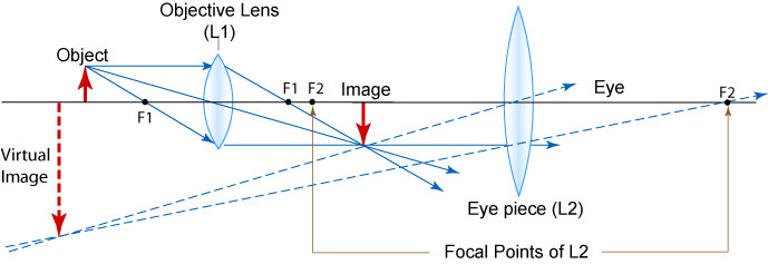
The use of microscopes help us to get magnified images, but how does it do that ? Actually f you remember , the microscope has 2 lenses, the objective lens and the optical lens. Boh these lenses act together to create a virtual image of the object , that is much larger than the original one. A look at the ray diagram will hell you clear the idea.
STAINING OF CELLS
IN ORder that the cells may be visible under the microscope with all the necessary details up to a certain magnification, we need to color them with specific dyes that change the transparency of the specimen and blocks the photons from the light source to penetrating the transparent specimen. That produces a differential dark and lighted area corresponding to the organelles that has taken absorbed the stain. The stain is is the compound that is obtained either chemically or can be obtained from natural sources like haematoxylin is obtained from the plant Caesalpinia spinosa and there are different types of hematoxylin, like Mayers Hematoxylin, Delafield’s HEmatoxylin etc. However , haematoxylin cannot be used directly as it is not actually a dye but has to be ripened (or oxidized to hematin ) by different processes.The section after bluing is counter stained with eosin.
MICROSCOPY
THE PROPER WAY OF USING THE MICROSCOPE IN LAB
COMPOUND MICROSCOPE
HOW DO YOU GIVE A LOOK AT THE PROKARYOTES?
Well as the name signifies, since they are too small to be seen by the naked eye, we need the microscope .The microscopes can be of many types , ranging from the simple to the complex and the Electron microscopes. However,let’ see how it is to be used

